







Uric acid circulates through the human bloodstream as the final byproduct of purine breakdown, a routine metabolic cleanup of DNA, RNA, and cellular fuel. While most mammals produce an active enzyme called uricase that degrades uric acid into water-soluble allantoin, human ancestors lost this genetic capability roughly 15 million years ago. As a result, humans live near the edge of chemical saturation. When serum urate levels cross 6.8 milligrams per deciliter (mg/dL), the bloodstream can no longer keep the compound dissolved. It precipitates out of solution as needle-sharp monosodium urate crystals, triggering explosive joint inflammation, renal stones, and systemic vascular strain.
Roughly 70 percent of daily uric acid leaves the body through renal filtration, while the remaining 30 percent exits through the gastrointestinal tract. When this filtration balance fails, uric acid accumulates rapidly. Clinical data shows that nearly 90 percent of hyperuricemia cases stem from renal underexcretion rather than excessive production. Yet the consequences reach far beyond sudden joint pain, quietly damaging blood vessels, raising blood pressure, and taxing metabolic health.
The 6.8 mg/dL Threshold: The Physics of Crystal Formation
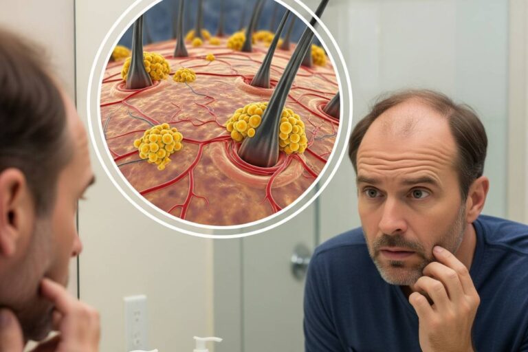
When serum urate surpasses 6.8 mg/dL (404 µmol/L) at normal body temperature, the blood becomes supersaturated. Urate molecules begin clumping together into microscopic, needle-shaped monosodium urate crystals.
Peripheral joints—especially the base of the big toe, known clinically as the first metatarsophalangeal joint—run slightly cooler than the core body. This lower temperature drops urate solubility further, making the feet and ankles prime targets for crystal deposition. Once these jagged crystals settle into synovial fluid between joints, the immune system treats them as foreign invaders.
Resident macrophages engulf the crystals and activate the NLRP3 inflammasome, an intracellular molecular alarm. This trigger unleashes a wave of interleukin-1 beta (IL-1β), a potent inflammatory cytokine. The result is acute gouty arthritis, marked by sudden nocturnal swelling, burning heat, and pain so severe that even the touch of a bedsheet feels unbearable. If hyperuricemia persists untreated for years, urate deposits solidify into chalky, granulomatous clusters called tophi. These tophi erode bone, destroy cartilage, and deform hands, elbows, and ears.
The Filtration Bottleneck: Why Urate Levels Rise
Elevated uric acid stems from two distinct physiological breakdowns: impaired kidney excretion or metabolic overproduction.
Renal Underexcretion Accounts for 90% of Cases
The kidneys filter urate through the glomeruli and actively reabsorb and secrete it along the proximal tubules using dedicated transport proteins, primarily URAT1 and GLUT9. When kidney function declines, urate clearance drops immediately.
Other circulating compounds also compete for these same tubular transporters. Elevated lactic acid from alcohol consumption and ketone bodies produced during strict fasting or ketogenic diets block uric acid secretion. Common medications—notably loop and thiazide diuretics prescribed for hypertension—similarly restrict urate clearance in the renal tubules, inadvertently driving up serum concentrations.
Metabolic Overproduction and Dietary Triggers
Approximately 10 percent of hyperuricemia cases arise from an oversupply of purines entering the bloodstream. Rapid cell turnover accelerates purine release in conditions such as psoriasis, hemolytic anemia, and tumor lysis syndrome following cancer chemotherapy.
Dietary inputs also intensify hepatic purine catabolism:
- Purine-Rich Foods: Organ meats, sardines, anchovies, and shellfish deliver concentrated loads of adenine and guanine, stressing hepatic clearance capacity.
- Fructose Overload: Unlike glucose, dietary fructose consumed in sweetened beverages and high-fructose corn syrup undergoes rapid phosphorylation by the hepatic enzyme fructokinase. This rapid reaction depletes cellular adenosine triphosphate (ATP), converting it to adenosine monophosphate (AMP). The sudden surge of AMP enters the purine breakdown cascade, driving rapid spikes in uric acid synthesis.
The Systemic Footprint: Beyond Arthritic Joints
Uric acid was long regarded solely as a rheumatologic problem. Modern vascular cardiology reveals that soluble, intracellular uric acid acts as a pro-inflammatory agent throughout the circulatory system.
Within vascular endothelial cells, intracellular urate blunts endothelial nitric oxide synthase (eNOS), reducing nitric oxide production. Without sufficient nitric oxide, blood vessels lose their ability to dilate smoothly, prompting arterial stiffness and sustained hypertension. At the same time, urate stimulates vascular smooth muscle proliferation and triggers the renin-angiotensin-aldosterone system (RAAS), accelerating renal arteriolosclerosis and raising cardiovascular risk.
In the kidneys, low urinary pH (falling below 5.5) dramatically reduces urate solubility in the collecting ducts. Free uric acid crystallizes into solid stones, accounting for roughly 10 percent of all kidney stone diagnoses and causing renal colic, urinary tract obstruction, and hematuria.
| Clinical Condition | Pathological Mechanism | Clinical Presentation |
|---|---|---|
| Acute Gouty Arthritis | Monosodium urate crystal precipitation in synovial fluid activating the NLRP3 inflammasome. | Sudden, severe joint pain, erythema, and heat (most commonly in the first metatarsophalangeal joint). |
| Chronic Tophaceous Gout | Persistent hyperuricemia forming granulomatous deposits of urate crystals and foreign-body giant cells. | Chalky subcutaneous nodules (tophi) causing irreversible joint deformities in hands, feet, and ears. |
| Uric Acid Nephrolithiasis | Low urinary pH (<5.5) prompting crystal aggregation in renal collecting ducts. | Severe flank pain, blood in urine, hydronephrosis, and recurrent urinary infections. |
| Endothelial & Vascular Strain | Intracellular urate inhibiting eNOS and stimulating vascular smooth muscle proliferation. | Arterial stiffness, persistent essential hypertension, and increased cardiometabolic strain. |
Clinical Benchmarks and Therapeutic Targets
Routine reference ranges define normal serum uric acid between 3.4 and 7.0 mg/dL for men, and between 2.4 and 6.0 mg/dL for premenopausal women, whose higher estrogen levels naturally promote renal urate clearance.
However, standard reference ranges do not apply once gout manifests. According to clinical guidelines established by the American College of Rheumatology (ACR), therapeutic management requires lowering serum urate well below the 6.8 mg/dL saturation threshold:
- Target for Standard Gout: Maintain serum urate below 6.0 mg/dL (360 µmol/L) to prevent crystal formation.
- Target for Severe or Tophaceous Gout: Lower serum urate below 5.0 mg/dL (300 µmol/L) to accelerate the physical dissolution of existing crystal stores and shrink tophi.
First-line medical therapy relies on Xanthine Oxidase Inhibitors (XOIs), such as allopurinol and febuxostat. These agents block the molybdenum-dependent enzyme xanthine oxidase from converting hypoxanthine and xanthine into uric acid. For patients who under-excrete urate despite healthy kidneys, uricosuric agents like probenecid inhibit renal URAT1 transporters to boost urinary excretion.
Practical Strategies for Urate Management
Pharmaceutical therapy remains the cornerstone for dissolving existing crystal deposits, but targeted dietary and metabolic adjustments significantly reduce hepatic burden and ease kidney workload.
Eliminating sugar-sweetened beverages, moderating alcohol intake (especially beer, which contains both alcohol and brewer’s yeast purines), and staying well hydrated keep urine volume high and lower crystallization risk. Those seeking specific meal planning strategies can explore our comprehensive uric acid diet guide: foods to eat and avoid, or examine the hydration and alkalizing effects covered in our review of lemon water for uric acid management.
Tracking serum urate through periodic blood testing enables clinicians and patients to catch hyperuricemia before permanent joint erosion or vascular damage takes hold.