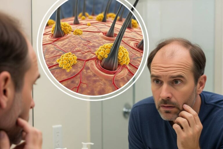







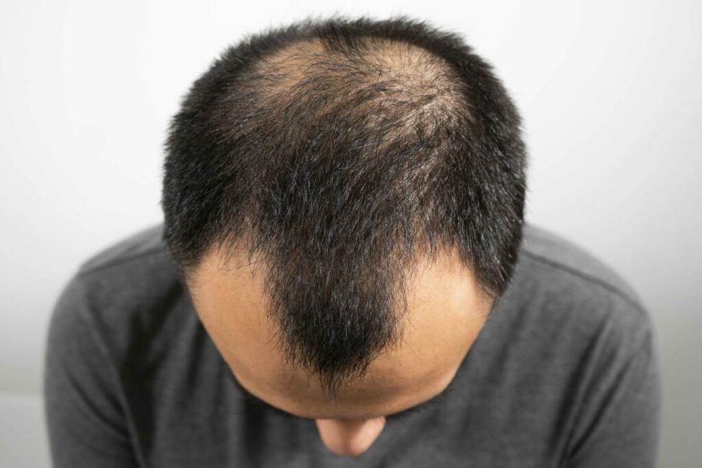
Pattern hair loss is not caused by an excess of circulating hormones, but by an inherited vulnerability where hair follicles interpret normal androgen signals as an instruction to deteriorate. Known medically as androgenetic alopecia, the condition affects an estimated 50 million men and 30 million women in the United States. Rather than triggering sudden shedding, it drives a slow structural decline known as follicular miniaturization, gradually shrinking robust terminal hairs into wispy, barely visible fibers over successive growth cycles.
The Cellular Mechanism: How DHT Chokes Follicle Growth
Androgenetic alopecia is a polygenic condition dictated by variations across multiple chromosomes, including the androgen receptor gene on the X chromosome and autosomal loci on chromosomes 3 and 20. The primary biochemical driver is dihydrotestosterone (DHT), a potent androgen synthesized locally in scalp tissue when the enzyme 5-alpha reductase metabolizes circulating testosterone. As documented in clinical genetics evaluations archived on PubMed Central, the severity of hair loss corresponds directly to follicular androgen receptor sensitivity rather than systemic testosterone levels.
When DHT binds to receptors within the follicular dermal papilla, it initiates an adverse signaling cascade. It stimulates the release of inhibitory cytokines, notably transforming growth factor-beta 1 (TGF-β1) and interleukin-6, which prematurely halt the anagen (growth) phase. A healthy scalp hair grows continuously for three to seven years. In vulnerable follicles exposed to DHT, this window contracts to months or weeks, while the telogen (resting) phase expands. With each aborted cycle, the dermal papilla shrinks in volume, anchors shallower in the dermis, and produces a thinner, unpigmented hair shaft until the follicle ceases production entirely.
Diverging Phenotypes: Norwood Versus Ludwig Progression
While the underlying endocrine process remains consistent across sexes, the anatomical manifestation differs sharply between men and women due to distinct enzyme densities across scalp zones.
In men, 5-alpha reductase activity and androgen receptor concentrations cluster heavily along the frontotemporal hairline and the vertex. This localized distribution yields the classic Hamilton-Norwood progression, beginning with symmetrical temple recession and crown thinning before converging into extensive baldness. The occipital rim along the back and sides of the head maintains minimal androgen receptor density, leaving those follicles genetically protected against DHT. For a deeper breakdown of masculine hormonal dynamics, see our clinical review on causes and treatments for hairfall in men.
Women experience a fundamentally different structural presentation categorized by the Ludwig and Savin scales. Female pattern hair loss rarely produces complete bald spots or a receding frontal hairline. Instead, higher scalp concentrations of the aromatase enzyme—which converts androgens into estrogens—protect the frontal boundary. Miniaturization occurs diffusely across the crown and central part, often presenting as a characteristic ‘Christmas tree’ distribution that widens the visible scalp line. Understanding these hormonal shifts is critical during life stages like perimenopause; read our guide to female hairfall and hormonal transitions for targeted guidance.
Systemic Biomarkers and Compounding Scalp Stressors
Dermatologists increasingly assess androgenetic alopecia within a broader systemic framework. Early-onset, aggressive miniaturization frequently correlates with metabolic and cardiovascular risk factors, including insulin resistance and dyslipidemia. Emerging data exploring how cholesterol metabolism impacts follicular microvasculature reveals that microcirculatory compromise can accelerate follicle starvation.
Accurate diagnosis also requires ruling out superimposed diffuse shedding. Telogen effluvium triggered by systemic illness, severe stress, or nutritional deficiencies such as low ferritin and vitamin D can dramatically accelerate the visual appearance of pattern baldness. Correcting these systemic deficits stabilizes shedding, allowing clinicians to isolate and treat the underlying genetic miniaturization.
Clinical Interventions: Halting Miniaturization Before Fibrosis
Pharmaceutical management must begin before miniaturized follicles undergo perifollicular fibrosis—a permanent scarring process that destroys the follicle stem cell niche. Therapeutic protocols outlined in our analysis of medical options for alopecia center on two primary objectives: suppressing DHT synthesis and stimulating microvascular perfusion.
- 5-Alpha Reductase Inhibitors: Oral finasteride selectively blocks the Type II isoenzyme, lowering scalp and serum DHT by roughly 70 percent to halt progression in over 80 percent of male patients. Oral dutasteride inhibits both Type I and Type II isoenzymes, reducing serum DHT by more than 90 percent for resistant cases.
- Vasodilators and Potassium Channel Openers: Minoxidil, applied as a 5 percent topical solution or prescribed as low-dose oral therapy (1.25 to 2.5 mg daily), upregulates vascular endothelial growth factor (VEGF) and opens potassium channels, forcing dormant follicles into an extended anagen phase.
- Endocrine Modulators for Women: Because 5-alpha reductase inhibitors carry teratogenic risks for women of childbearing potential, female patients often benefit from competitive androgen receptor antagonists such as spironolactone or cyproterone acetate under specialist supervision.
- Surgical Autologous Transplantation: Follicular Unit Extraction (FUE) and Follicular Unit Transplantation (FUT) surgically harvest androgen-resistant donor follicles from the occipital scalp and relocate them to miniaturized regions, where they permanently retain their original DHT immunity.
The Therapeutic Window: Why Early Action Dictates Outcomes
Androgenetic alopecia is a progressive biological clock. Because pharmaceutical agents cannot resurrect follicles that have fully fibrosed and atrophied, clinical success depends almost entirely on baseline preservation. Patients who initiate targeted medical therapy at the first sign of temple recession or part-line widening retain substantially more follicular density over decades than those who delay until scalp skin becomes visible.