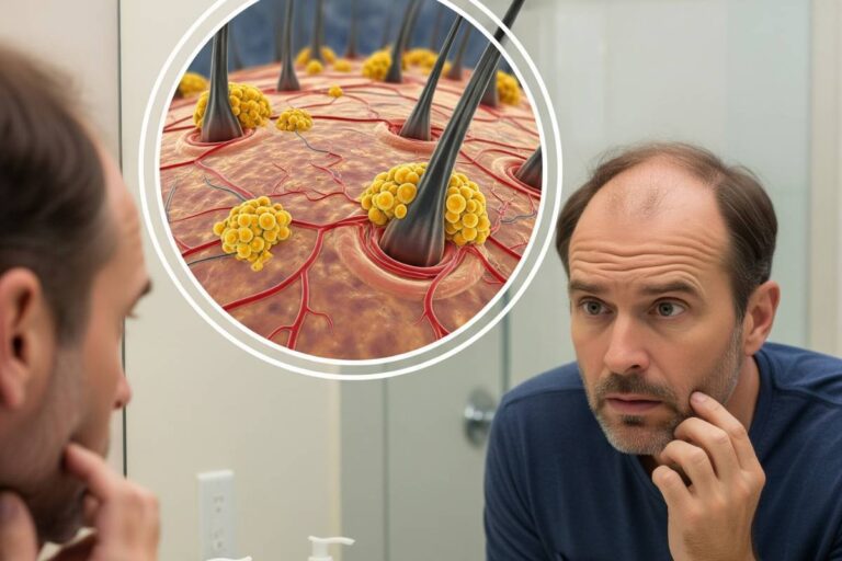








Androgenetic alopecia, commonly recognized as male pattern hair loss, affects roughly 30 percent of men by age 30 and more than 50 percent by age 50. Contrary to widespread myths attributing thinning hair to frequent shampooing, headwear, or surface blood flow, the condition is an inherited, progressive endocrine disorder. The biological driver is a heightened sensitivity in scalp follicles to dihydrotestosterone (DHT), a potent derivative of testosterone. Without medical intervention, susceptible follicles shrink over successive growth cycles until they cease producing terminal hair fibers altogether.
Clinical management has shifted decisively toward early, targeted intervention. Landmark research published in the Journal of the American Academy of Dermatology confirms that medical stabilization yields significantly higher follicular density when initiated during early thinning rather than after extensive follicular scarring has occurred. Halting progression requires understanding the underlying hormonal cascade, separating androgenic loss from acute telogen shedding, and deploying evidence-based therapeutic regimens.
The Biology of Follicular Miniaturization: DHT and Androgen Receptors
Male pattern hair loss centers on an enzymatic conversion taking place directly within the scalp tissue. The enzyme Type II 5-alpha reductase converts circulating testosterone into dihydrotestosterone (DHT). While DHT is critical for male fetal development and secondary sexual characteristics during puberty, its post-pubertal activity in the scalp triggers progressive follicular regression.
Follicular vulnerability follows a strict anatomical distribution. Hair follicles on the temples, crown, and vertex possess dense concentrations of androgen receptors and high levels of 5-alpha reductase enzymes. In contrast, follicles located along the occipital rim at the back and lower sides of the scalp carry few androgen receptors and remain genetically resistant to DHT. Detailed clinical reviews of androgenetic alopecia mechanisms show how this hormonal exposure systematically alters the hair cycle:
- Shortened Anagen Phase: The active growth phase drops from a normal duration of two to six years down to several months or weeks.
- Prolonged Kenogen Phase: The latency period between the shedding of an old hair and the emergence of a new shaft lengthens substantially, leaving follicles empty for longer spans.
- Progressive Miniaturization: Each shortened cycle yields a thinner, shorter, and less pigmented shaft, eventually reducing robust terminal hairs into microscopic, vellus-like fuzz.
- Irreversible Fibrosis: If miniaturization continues unchecked over years, the dermal papilla detaches, and connective tissue scarring replaces the follicle, eliminating any potential for non-surgical regrowth.
Distinguishing Pattern Thinning from Acute Shedding and Lifestyle Triggers
While androgen sensitivity dictates whether a man develops pattern baldness, several secondary physiological insults can accelerate the rate of follicular shedding. Accurately diagnosing the primary cause prevents misdirected treatments.
Acute telogen effluvium represents the most frequent non-genetic contributor. Under intense physical trauma, severe psychological crises, or prolonged systemic illness, high circulating levels of cortisol shock up to 30 percent of actively growing hairs into a premature resting phase. Within two to three months of the inciting event, diffuse shedding occurs across the entire scalp rather than following the distinct temple and vertex recession characteristic of androgenetic alopecia. Clinical assessments of how systemic stress alters physical health illustrate that resolving the underlying stressor typically restores baseline growth within six to nine months, provided the follicles are not already compromised by DHT.
Nutritional deficiencies and microvascular constriction also compromise cellular turnover at the follicular base:
- Depleted Iron and Ferritin Stores: Serum ferritin levels below 40 ng/mL restrict oxygen delivery to rapidly dividing follicular matrix cells. Evaluation of vitamin deficiencies and hair shedding reveals that correcting low iron, vitamin D, and zinc levels is essential for optimal hair shaft synthesis.
- Microvascular Damage from Smoking: Nicotine promotes acute vasoconstriction within the delicate capillary networks feeding the dermal papilla. Long-term observational studies show that men who smoke experience higher rates of accelerated thinning compared to non-smokers due to elevated oxidative stress and compromised microvascular circulation.
- Seborrheic Dermatitis and Scalp Inflammation: Chronic microbial overgrowth of Malassezia yeast generates localized inflammation and oxidative stress, which aggravates follicular miniaturization and accelerates shed rates in predisposed men.
Evidence-Based Medical Therapies: Clinical Protocols and Timelines
Dermatologists rely on therapeutic protocols designed either to inhibit androgen conversion or to stimulate microvascular follicular perfusion. Combining these distinct mechanisms yields higher rates of stabilization and regrowth than monotherapy alone.
5-Alpha Reductase Inhibitors: Halting Hormonal Miniaturization
Oral finasteride at a dosage of 1 milligram daily remains the frontline medical treatment for stabilizing male pattern thinning. The medication competitively inhibits the Type II 5-alpha reductase enzyme, reducing circulating serum DHT levels by approximately 70 percent and scalp tissue DHT by roughly 65 percent. Long-term multi-center clinical trials demonstrate that oral finasteride halts hair loss progression in 85 to 90 percent of men over five years, with approximately 65 percent experiencing visible hair regrowth along the vertex.
For patients concerned about systemic absorption or rare side effects, compounding dermatologists increasingly prescribe topical finasteride solutions (typically 0.1% to 0.25%). Clinical data show topical formulations suppress scalp DHT locally while reducing systemic DHT inhibition to under 30 percent, offering a viable alternative with a lower risk of systemic adverse effects.
Microvascular and Cellular Stimulators: Minoxidil Regimens
Minoxidil operates through a distinct, non-hormonal pathway. As a potassium channel opener and vasodilator, minoxidil widens microvessels around the follicle, enhances local tissue perfusion, and upregulates vascular endothelial growth factor (VEGF). This mechanism shifts telogen-phase hairs into active anagen growth and extends the duration of follicular expansion. Patients initiating topical 5% minoxidil often observe a temporary shedding surge between weeks two and eight. This reaction represents a positive clinical sign indicating synchronized entry of dormant follicles into active growth.
In addition to topical foam and liquid formulations, dermatologists increasingly use low-dose oral minoxidil (1.25 mg to 2.5 mg daily) for men who experience scalp irritation or low compliance with twice-daily topical applications. Comprehensive breakdowns of medical treatments for alopecia demonstrate that combining finasteride with minoxidil achieves synergistic follicular defense.
| Treatment Modality | Primary Mechanism | Clinical Response Rate | Expected Timeline |
|---|---|---|---|
| Oral Finasteride (1 mg/day) | Type II 5-AR inhibitor; lowers scalp DHT by ~65% | Stops loss in 85–90%; regrowth in 65% | 3 to 6 months for stabilization; 12 months for peak density |
| Topical Minoxidil (5%) | Potassium channel opener; prolongs anagen growth | Stabilization and regrowth in 60–70% | Initial shedding at 4–8 weeks; visual gains at 4 to 6 months |
| Platelet-Rich Plasma (PRP) | Autologous growth factors stimulate dermal papilla | Variable; effective as an adjunct therapy | Requires 3 monthly loading sessions followed by maintenance |
| Follicular Unit Extraction (FUE) | Autologous graft relocation of DHT-resistant occipital follicles | Over 90% graft survival in experienced surgical centers | Transplanted shedding at 1 month; mature growth at 9 to 12 months |
Clinical Diagnostics and Physician Evaluation
Dermatologists classify the progression of male pattern thinning using the standardized Norwood-Hamilton scale, which ranges from Stage I (minimal frontoparietal recession) to Stage VII (severe loss leaving only an occipital fringe). Formal evaluation includes trichoscopic analysis—a non-invasive digital dermatoscopy technique that magnifies the scalp up to 70 times.
Under trichoscopy, a diagnosis of androgenetic alopecia is confirmed when follicle diameter diversity exceeds 20 percent across the frontal or crown scalp, accompanied by peripilar halos (perifollicular pigmentation signifying ongoing micro-inflammation). When hair loss patterns present atypically, physicians perform a 4-millimeter punch biopsy or targeted blood panels checking ferritin, total testosterone, free testosterone, and thyroid-stimulating hormone (TSH). To understand diagnostic procedures in detail, review how clinicians diagnose alopecia. Patients considering non-pharmaceutical options should examine clinical assessments of botanical and dietary adjuncts for hair health to separate evidence-supported supplements from commercial marketing.
Long-Term Management and Treatment Trajectories
Maintaining follicular density over decades requires an understanding of treatment continuity. Because androgenetic alopecia is genetically preprogrammed, medical treatments do not permanently alter follicle genetics. Discontinuing finasteride or minoxidil leads to a gradual reversal of therapeutic gains within six to twelve months, returning the scalp to the baseline level of thinning that would have developed without treatment.
Surgical restoration via Follicular Unit Extraction (FUE) provides durable hairline reconstruction because occipital donor follicles retain their genetic resistance to DHT even after transplantation. However, without concurrent medical therapy to protect non-transplanted native follicles behind the graft zone, thinning will continue unabated around the transplanted hair. Men observing early temporal recession or vertex thinning achieve the greatest long-term density by scheduling a trichoscopic assessment with a board-certified dermatologist and establishing an evidence-based maintenance regimen before permanent follicular scarring sets in.