






An accurate alopecia diagnosis separates temporary, reversible shedding from permanent follicular destruction. While hair thinning often looks identical on the surface, dermatologists rely on a systematic diagnostic pipeline—combining medical chronology, non-invasive digital trichoscopy, targeted blood panels, and histopathologic tissue biopsy—to identify the precise condition among more than two dozen hair loss disorders. Because misdiagnosing scarring conditions like frontal fibrosing alopecia or lichen planopilaris can lead to irreversible follicular loss within months, clinical protocols prioritize definitive differential diagnosis before selecting medical therapies.
Clinical Chronology: Mapping Shedding Velocity and Scalp Symptoms
Diagnostic evaluation begins with a meticulous clinical history to chart shedding velocity, distribution, and precipitating triggers. Dermatologists categorize hair loss by its speed of onset:
- Acute Diffuse Shedding: An abrupt loss of hundreds of hairs daily typically indicates acute telogen effluvium or rapid-onset alopecia areata. Because telogen effluvium operates on a predictable two-to-four-month physiological lag following a trigger, clinicians investigate major stressors such as severe infections, high fevers, childbirth, surgical procedures, or rapid nutritional deficits.
- Insidious Progressive Thinning: Gradual thinning that develops over months or years across the crown, temples, or frontal hairline points toward androgenetic alopecia (pattern hair loss).
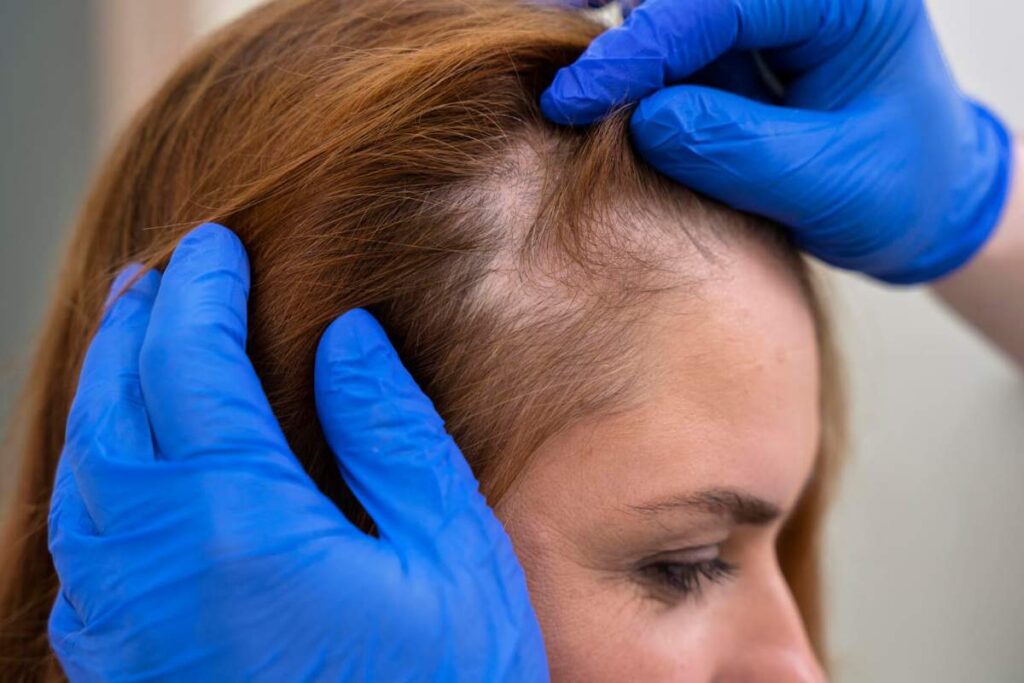
- Patchy or Circumscribed Loss: Rapid focal hair shedding presenting as smooth circular patches indicates autoimmune alopecia areata, while expanding hairless patches accompanied by skin surface changes raise immediate suspicion of primary cicatricial (scarring) alopecia.
Physicians also evaluate localized neurosensory symptoms. Pruritus (itching), burning sensations, or scalp tenderness (trichodynia) frequently signal active follicular inflammation in scarring conditions. Investigating family history, medication changes, and hormonal milestones helps clinicians map systemic contributors, as explored in our guide on types of alopecia explained.
Bedside Physical Examination: Standardized Hair Pull and Tug Tests
Before using specialized imaging devices, dermatologists perform standardized physical manipulation tests at the bedside to quantify active shedding and hair shaft fragility.
The standardized hair pull test evaluates active shedding across different scalp zones. The clinician grasps a bundle of approximately 50 to 60 terminal hairs between the thumb, index, and middle fingers near the scalp base and exerts gentle, continuous traction as the fingers slide along the shafts to the distal ends. A pull test is considered positive when more than 10% of grasped hairs (typically six or more) detach easily without pain. Positive results across multiple scalp locations indicate diffuse conditions like active telogen effluvium, whereas positive results localized to the periphery of bare patches point to active, expanding margins in alopecia areata.
Clinicians contrast the pull test with the hair tug test. By holding a hair shaft firmly near the scalp with one hand while pulling the distal shaft with the other, the physician applies isolated mechanical tension to the hair fiber. If the shaft snaps mid-strand, it confirms structural shaft fragility—such as trichorrhexis nodosa or genetic shaft defects—rather than an active root-level shedding disorder.
Digital Trichoscopy: Microscopic Follicular Patterns
Non-invasive dermatoscopy of the hair and scalp, known as trichoscopy, has transformed clinical practice by reducing the need for invasive biopsies. Using polarized light at 10x to 70x magnification, clinicians examine follicular openings (ostia), peripilar skin, hair shaft calibers, and microvascular networks. Guidelines published in the Journal of the American Academy of Dermatology identify distinctive micro-phenotypic criteria for common scalp disorders:
| Condition | Primary Trichoscopic Hallmarks | Pathophysiological Mechanism |
|---|---|---|
| Alopecia Areata | Exclamation mark hairs, black dots, yellow dots, micro-exclamation hairs | Peribulbar lymphocytic inflammation weakens the proximal shaft, causing structural fracture and follicular arrest. |
| Androgenetic Alopecia | Hair diameter diversity (>20%), peripilar brown halos, empty ostia | Progressive 5-alpha reductase-driven miniaturization converts robust terminal follicles into fine vellus hairs. |
| Telogen Effluvium | Preserved follicular openings, upright regrowing hairs, lack of diameter diversity | Premature transition of anagen follicles into the resting telogen phase without structural follicle shrinkage. |
| Cicatricial (Scarring) Alopecia | Complete loss of follicular ostia, peripilar casts, erythema, tubular scales | Permanent inflammatory destruction of follicular stem cells in the bulge, replaced by fibrous connective tissue. |
Trichoscopy distinguishes between non-scarring and scarring alopecias in seconds. While non-scarring forms maintain visible follicular openings containing single or grouped hairs, cicatricial conditions erase these openings entirely, leaving smooth, ivory-white scar tissue. Identifying peripilar casts—hyperkeratotic collars surrounding the emerging hair shaft—allows clinicians to detect lichen planopilaris or frontal fibrosing alopecia before irreversible bald patches form.
Targeted Blood Biomarkers: Ruling Out Metabolic and Endocrine Drivers
Hair matrix cells rank among the fastest-dividing cell populations in the human body, demanding substantial metabolic energy, micronutrients, and hormonal balance. Rather than ordering indiscriminate panels, dermatologists use targeted serological assays to uncover systemic drivers of diffuse thinning:
- Serum Ferritin: Reflects intracellular iron reserves. Hair follicle matrix proliferation requires iron-dependent ribonucleotide reductase activity. While standard reference laboratories report levels above 15 ng/mL as normal, trichology guidelines establish that serum ferritin below 50 ng/mL strongly correlates with persistent telogen effluvium and impaired anagen maintenance. Our detailed review of hair loss and vitamin deficiencies covers these nutritional thresholds in depth.
- Comprehensive Thyroid Panel (TSH, Free T3, Free T4): Both hypothyroidism and hyperthyroidism disrupt normal hair cycling, blunting mitotic division in the matrix and causing diffuse telogen conversion, brittle hair shafts, and outer third eyebrow thinning.
- 25-Hydroxyvitamin D: Vitamin D receptors in keratinocytes govern the normal progression of the hair cycle. Serum levels falling below 30 ng/mL compromise follicular cycling and delay anagen initiation.
- Endocrine and Androgen Profiling: Total and free testosterone, dehydroepiandrosterone sulfate (DHEA-S), and androstenedione clarify suspected hyperandrogenism. This assessment proves particularly valuable when evaluating hair loss in females with suspected PCOS or hormonal imbalances who present with pattern thinning alongside hirsutism or acne.
- Autoimmune Serology: Antinuclear antibodies (ANA) screen for systemic lupus erythematosus, which can cause both non-scarring telogen shedding and scarring discoid lupus lesions.
Histopathology: Scalp Punch Biopsy and the Headington Technique
When trichoscopy and clinical evaluations show equivocal findings—or when signs of scarring alopecia emerge—a scalp punch biopsy serves as the definitive gold standard. Dermatologists perform this minor surgical procedure under local anesthesia (typically 1% or 2% lidocaine with epinephrine), extracting a 4-millimeter full-thickness cylinder of scalp tissue that extends down into the subcutaneous fat layer.
Precise site selection remains critical for accurate pathology. The clinician harvests the biopsy specimen from an active, hair-bearing border displaying inflammatory signs rather than the center of an inactive, burned-out bald patch. The tissue sample is closed with one or two simple nylon sutures, producing a minor, concealed scar that heals within 7 to 10 days.
Modern dermatopathology relies on the Headington technique, utilizing both horizontal (transverse) and vertical tissue sectioning to maximize diagnostic yield:
- Horizontal (Transverse) Sections: Allow pathologists to evaluate all follicular units in a single field. Pathologists calculate the total hair count, determine follicular density per square millimeter, and measure the terminal-to-vellus hair ratio. In a healthy scalp, terminal hairs outnumber fine vellus hairs by approximately 7:1 to 8:1; in androgenetic alopecia, that ratio falls below 4:1 or 2:1. Transverse sections also quantify the percentage of follicles in catagen and telogen phases, confirming acute or chronic effluvium when telogen counts exceed 15% to 20%.
- Vertical Sections: Provide a panoramic longitudinal view from the epidermis down to the subcutaneous fat. This view identifies interface dermatitis, basement membrane thickening, apoptotic keratinocytes, and the precise depth of inflammatory cell infiltrates. While alopecia areata shows dense lymphocytic infiltration concentrated around the deep hair bulb (‘swarm of bees’), lichen planopilaris shows lichenoid lymphocytic inflammation centered higher along the infundibulum and isthmus.
Directing Clinical Therapy Through Diagnostic Clarity
Differentiating hair loss subtypes is not an academic exercise—it dictates medical strategy. Prescribing topical growth stimulators or vasodilators to an untreated patient with active scarring alopecia allows progressive inflammatory destruction to extinguish remaining follicular stem cells. Conversely, identifying active peribulbar autoimmune inflammation in alopecia areata directs clinicians toward localized anti-inflammatory therapies or systemic Janus kinase (JAK) inhibitors.
Once clinicians establish the underlying pathology and confirm whether follicles remain viable, they design targeted, evidence-based regimens. To evaluate modern pharmacologic approaches, review our clinical guide on medical treatments for alopecia. Prompt, objective diagnosis protects scalp hair density by replacing empirical guesswork with targeted clinical care.