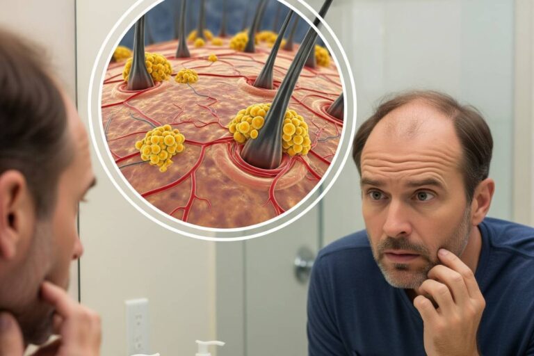







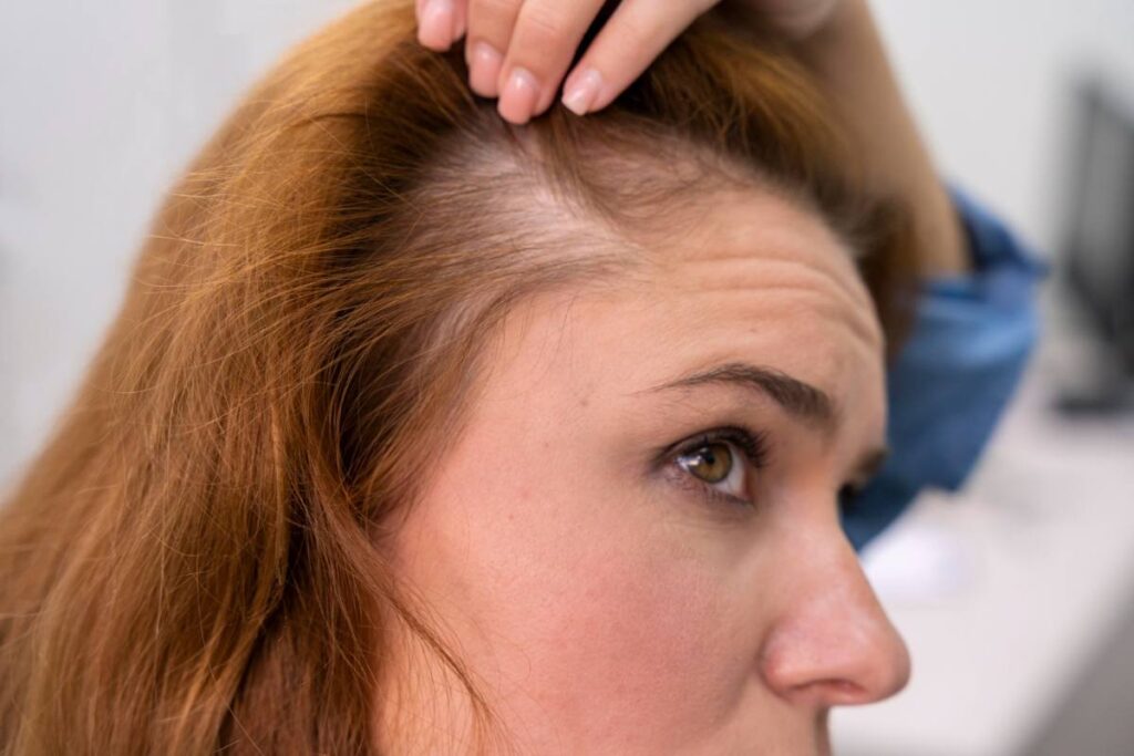
Alopecia is not a monolithic condition, but an expansive medical umbrella covering distinct immunological, hormonal, and mechanical disorders of the hair follicle. While common parlance often treats all hair loss as cosmetic thinning, dermatological prognosis hinges on a single biological boundary: follicular stem cell viability. In non-scarring forms, the follicle remains biologically active beneath the scalp, preserving the capacity for regrowth. In scarring (cicatricial) variants, destructive inflammation obliterates these regenerative reservoirs, permanently replacing hair-bearing tissue with fibrous scars.
Every clinical presentation demands rapid diagnostic precision. Because therapeutic pathways range from targeted immunomodulators to hormonal receptor blockers, distinguishing between temporary shedding and active follicular destruction determines whether hair density can be recovered or merely stabilized.
Follicular Cycling and the Stem Cell Architecture
Human hair growth operates through continuous, asynchronous cycles governed by complex biochemical signaling between the dermal papilla and epithelial stem cells located in the follicle bulge. At any given moment, the human scalp hosts roughly 100,000 follicles cycling through three distinct phases:
- Anagen (Active Growth): Spanning two to seven years, this phase defines the ultimate length of the hair shaft. Roughly 85% to 90% of scalp follicles remain in anagen under healthy conditions, driven by rapid cell division in the hair matrix.
- Catagen (Follicular Regression): Lasting two to three weeks, catagen marks an orderly apoptosis-driven involution. Cell proliferation halts, and the lower follicle regresses, pulling the hair shaft away from its primary subcutaneous blood supply.
- Telogen (Rest and Shedding): Over approximately 100 days, the hair club sits dormant while biochemical signals prepare the niche for a new growth cycle. The physiological shedding of 50 to 100 telogen hairs per day represents normal cellular turnover, not pathological loss.
When systemic illness, hormonal shifts, or immune cells disrupt these phase transitions, the synchronous loss of hairs manifests as clinical alopecia.
Non-Scarring Alopecia: Dormancy and Regrowth Potential
Non-scarring alopecias spare the follicular bulge stem cells. Although follicle diameter may diminish or hairs may shed en masse, the structural architecture necessary to generate a terminal hair remains intact.
Androgenetic Alopecia: Hormonal Miniaturization
Accounting for the vast majority of progressive thinning, androgenetic alopecia affects roughly 50% of men over age 50 and up to 40% of postmenopausal women. The disorder is driven by an inherited sensitivity of follicular androgen receptors to dihydrotestosterone (DHT), a potent androgen converted from circulating testosterone by the enzyme 5-alpha reductase.
DHT exposure causes progressive follicular miniaturization, shortening the anagen growth window while elongating the telogen phase. Over successive cycles, robust terminal hairs transform into wispy, unpigmented vellus hairs. In men, loss typically tracks the Hamilton-Norwood scale, starting with bitemporal recession and vertex thinning. In women, loss follows the Ludwig pattern, presenting as diffuse thinning along the central midline with preservation of the frontal hairline. Detailed clinical staging is explored in our breakdown of androgenetic alopecia male and female patterns.
Alopecia Areata: Collapsed Immune Privilege
Normal hair follicles maintain “immune privilege,” a specialized microenvironment that suppresses major histocompatibility complex (MHC) class I molecules to evade self-reactive immune surveillance. In alopecia areata, this protective shield collapses. Autoreactive CD8+ T lymphocytes cluster around the anagen hair bulb in a distinctive “swarm of bees” histopathological pattern, prematurely driving the hair into dystrophic catagen.
According to clinical data compiled by the National Alopecia Areata Foundation, the condition manifests along an unpredictable spectrum:
- Patchy Alopecia Areata: Circumscribed, smooth, round or oval patches of sudden hair loss on the scalp or beard, often accompanied by exclamation-point hairs visible under dermoscopy.
- Alopecia Totalis: Complete shedding across the entire scalp.
- Alopecia Universalis: Complete loss of terminal hair across the entire body, including eyebrows, eyelashes, and axillary hair.
While historically treated primarily with local corticosteroids, severe presentations are now managed using oral Janus kinase (JAK) inhibitors such as baricitinib and ritlecitinib, which interrupt intracellular cytokine signaling (interferon-gamma and interleukin-15) to restore immune privilege.
Telogen Effluvium: Diffuse Phase Synchrony
Unlike localized thinning, telogen effluvium is a reactive, diffuse loss pattern that emerges when an acute physiological or psychological shock precipitates widespread follicular transitions. Severe systemic infections, major surgery, rapid weight loss, high-grade fevers, and postpartum hormonal crashes can prematurely push up to 30% to 50% of growing anagen follicles into the telogen resting phase.
Because the telogen phase lasts approximately 90 to 120 days, shedding typically manifests two to three months after the triggering event. Patients frequently report dramatic hair loss during brushing or washing. Once the acute metabolic trigger resolves, follicular kinetics normalize, and full density generally returns within six to nine months.
Traction Alopecia: Mechanical Strain
Traction alopecia stems from sustained, mechanical tension exerted on hair roots by tight styling practices, including braids, cornrows, heavy dreadlocks, weaves, and tightly pulled ponytails. Chronic tension induces localized folliculitis around the hair margins, particularly along the temporal and frontal borders.
Early-stage traction alopecia is fully reversible once tension is eliminated. However, persistent traction over years provokes mechanical destruction and secondary perifollicular fibrosis, permanently destroying the follicle root and transitioning into scarring alopecia.
Cicatricial Alopecia: Dermatologic Emergencies
Scarring, or cicatricial, alopecias represent medical emergencies within dermatology, as outlined by the American Academy of Dermatology. In these conditions, inflammation targets the upper portion of the follicle—the bulge and infundibulum—where multipotent stem cells and sebaceous glands reside.
Once stem cells are destroyed, the follicle cannot regenerate a new hair shaft. The follicular ostium closes and is replaced by fibrotic connective tissue, creating smooth, shiny patches where pore openings are absent. Common variants include:
- Lichen Planopilaris (LPP): A T-cell-mediated autoimmune condition characterized by violaceous perifollicular erythema, follicular hyperkeratosis, and burning scalp pain.
- Frontal Fibrosing Alopecia (FFA): A progressive band-like recession of the frontal and temporal hair margins, predominantly affecting postmenopausal women and frequently accompanied by complete loss of eyebrow hair.
- Central Centrifugal Cicatricial Alopecia (CCCA): A progressive scarring condition that initiates at the vertex or crown and expands symmetrically outward, seen primarily in women of African descent.
Prompt clinical intervention with potent anti-inflammatory agents or immunomodulators aims to halt active peripheral expansion, though lost follicular architecture cannot be reconstructed.
Differential Diagnosis and Diagnostic Protocols
Accurate diagnosis prevents inappropriate treatments, such as applying topical vasodilators to active autoimmune or scarring conditions. Dermatologists employ a structured diagnostic framework:
- Trichoscopy: High-magnification polarized dermoscopy reveals micro-structural diagnostic markers: exclamation point hairs and yellow dots in alopecia areata; hair shaft thickness heterogeneity exceeding 20% in androgenetic alopecia; peripilar casts and erythema in cicatricial disease.
- Hair Pull Test: Gentle traction applied to clusters of 50 to 60 hairs assesses active shedding versus resting baseline. Yielding more than 10% telogen bulbs confirms active effluvium.
- Scalp Biopsy: A 4-mm punch biopsy provides cross-sectional (transverse) and vertical views under histopathology, definitively distinguishing between inflammatory cicatricial infiltrates and non-scarring follicular miniaturization. A complete clinical walk-through is detailed in our guide on how dermatologists diagnose alopecia with trichoscopy and biopsies.
- Serum Biomarker Screening: Systemic screens check for contributing metabolic drivers, including serum ferritin, thyroid-stimulating hormone (TSH), complete blood counts, and vitamin D levels. The biochemical role of these micronutrients is covered in our review of ferritin and vitamin D deficiencies triggering telogen effluvium.
Clinical Management Pathways
Therapeutic interventions must target the specific cellular mechanism driving follicle impairment:
- Follicular Miniaturization: Managed with topical or oral minoxidil to prolong anagen duration and improve microvascular perfusion, paired with oral 5-alpha reductase inhibitors (finasteride, dutasteride) to curb DHT production. An overview of these options is available in our analysis of medical treatments for hair regrowth.
- Immune Privilege Disruption: Treated with high-potency topical or intralesional triamcinolone acetonide injections for localized patches, or systemic JAK inhibitors for extensive alopecia totalis or universalis.
- Cicatricial Inflammation: Managed aggressively with topical calcineurin inhibitors, oral antimalarials (hydroxychloroquine), or oral tetracyclines to quench follicular inflammation before permanent fibrotic conversion.
The Clinical Imperative of Early Intervention
The trajectory of any hair loss diagnosis depends on the speed of clinical evaluation. While diffuse shedding often resolves following metabolic stabilization, progressive conditions like androgenetic alopecia require continuous suppression of hormonal triggers before miniaturized follicles undergo complete senescence. For scarring alopecias, every week of uncontrolled inflammation represents permanent follicular attrition. Consulting a board-certified dermatologist at the first sign of unexpected shedding, scalp tenderness, or receding margins remains the most reliable strategy to safeguard follicular health.